
A Diagnosis Without the Long Journey
For years, access to X-ray services at Chikankata Mission Hospital was uncertain. The old machine broke down frequently, often without warning. When it failed, patients waited. When it could not be repaired quickly, they traveled.
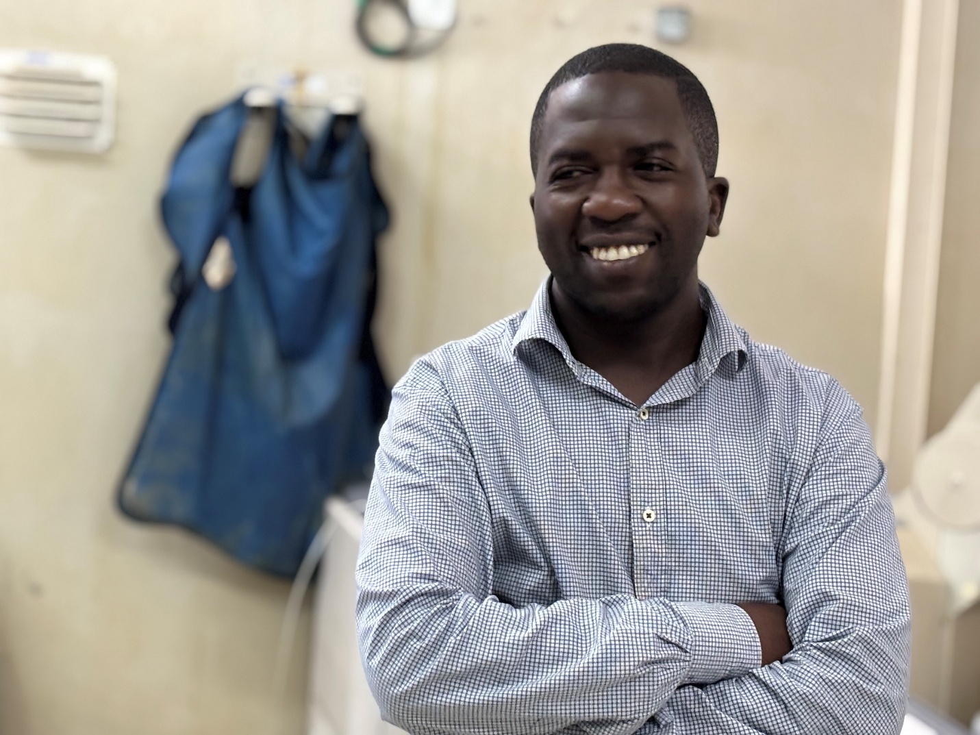
Eventually, the machine stopped working altogether. For more than a year, Chikankata had no functioning Xray equipment. Patients who needed imaging to confirm a fracture, assess an injury, or support treatment for tuberculosis were sent elsewhere. On an average day, at least 20 patients who required Xray imaging were referred out, often to Kafue or Lusaka, because diagnostic services were unavailable on site. As radiographer Mulwa Phiri explained, “that was the minimum number we were referring every day,” adding that many of those journeys were “very far and very costly for the patients.”
For patients already in pain, the journey itself was a burden. Many of those referrals were for fractures caused by road traffic and oxcart accidents, which make up the majority of Xray cases seen at the hospital. Others were for chest infections, including suspected tuberculosis, where treatment decisions depend on imaging results. For these patients, delays stretched from days into weeks, not because care was unavailable, but because diagnosis was far away.
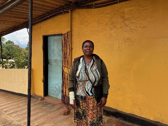
Shelly Mainza remembers that period clearly. In 2024, she injured her back. With no Xray services available at Chikankata, she traveled repeatedly to seek care elsewhere. Each trip cost money she could not easily replace. Diagnosis and treatment took time because of the back and forth required to get the x-rays she needed elsewhere.
When Shelly returned to Chikankata in 2026 with a broken hand, the experience was completely different. Thanks to funding support from the Helmsley Charitable Trust, the hospital now had a new digital Xray machine. Her Xray was taken on site, and the image was transmitted digitally. Her Xray was taken on site, and the image was transmitted digitally. By the time she walked around the corner to the physiotherapy unit, her X-rays were already there. The injury was confirmed the same day, and treatment began immediately. The entire process, from imaging to diagnosis, was provided at no cost, eliminating the travel, delay, and expense she had faced before.
For patients, the change is immediate and practical. Injuries no longer require long journeys just to confirm what is wrong. Suspected fractures can be assessed quickly. TB patients can be diagnosed and monitored without leaving the community. Care begins sooner, and unnecessary travel is avoided.
From the hospital’s perspective, the difference is consistency. Imaging is no longer dependent on whether an aging machine is working or whether patients can afford to travel. Results are available quickly, allowing clinicians to make decisions without delay and coordinate care more effectively.
While the new Xray machine does not replace clinical expertise, it does change how quickly diagnosis can occur and where it takes place. Patients can now be assessed and treated at Chikankata without traveling elsewhere or delaying care. This provides consistent access to diagnostic imaging for injuries and for conditions such as tuberculosis, supporting both emergency decisions and ongoing treatment within the hospital.
When diagnosis happens in time
At Chikankata, the most immediate impact of the new digital Xray machine has been on patients whose care depends on timely diagnosis, particularly those with fractures and those with suspected tuberculosis.
Before the new machine was installed, most patients needing Xray imaging were referred elsewhere. According to radiography staff, road traffic and oxcart accidents account for the majority of Xray cases at the hospital. These patients often arrived in pain, with injuries that could not be safely treated without imaging. When the old machine was not working, they were sent to distant facilities, delaying treatment and increasing the risk of complications.
With digital X-ray now available on site, those patients are assessed immediately. Fractures can be confirmed the same day, allowing clinicians to immobilize injuries, begin physiotherapy, or determine whether surgical referral is needed without delay. The difference is not technological sophistication, but timing: injuries are managed before swelling worsens, before patients attempt to travel, and before untreated fractures lead to longer-term disability.
Chest infections present a similar risk when diagnosis is delayed. For patients with suspected tuberculosis, Xray imaging is often necessary to confirm disease and guide treatment decisions. In the past, these patients were also referred out when imaging was unavailable. “Some of the management depends on the Xray,” radiographer Mulwa Phiri explained, meaning treatment could not move forward without it.
Today, those patients can be imaged at Chikankata. Diagnosis and follow-up no longer depend on travel, repeated visits, or waiting for results from other facilities. Treatment decisions are made sooner, reducing the risk that illness progresses while patients wait for confirmation.
In both cases, the impact is the same: care begins when patients arrive, often within a few minutes of arriving. The digital X-ray machine has removed a critical bottleneck in diagnosis, allowing injuries and serious infections to be identified and treated before they become life-threatening.